CD2
| editar |
| CD2 molecule | |||||||||||||
|---|---|---|---|---|---|---|---|---|---|---|---|---|---|
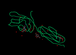 Estrutura proteica de CD2 a partir de PDB 1hnf | |||||||||||||
| |||||||||||||
| Identificadores | |||||||||||||
| Símbolos | CD2; LFA-2; SRBC; T11 | ||||||||||||
| IDs externos | OMIM: 186990 MGI: 88320 HomoloGene: 1338 ChEMBL: 2040 GeneCards: CD2 Gene | ||||||||||||
| |||||||||||||
| Padrões de expressão do ARN | |||||||||||||
 | |||||||||||||
| Mais dados de expressão | |||||||||||||
| Ortólogos | |||||||||||||
| Espécies | Humano | Rato | |||||||||||
| Entrez | 914 | 12481 | |||||||||||
| Ensembl | ENSG00000116824 | ENSMUSG00000027863 | |||||||||||
| UniProt | P06729 | P08920 | |||||||||||
| RefSeq (mRNA) | NM_001767 | NM_013486 | |||||||||||
| RefSeq (proteína) | NP_001758 | NP_038514 | |||||||||||
| Localização (UCSC) | Chr 1: 117.3 – 117.31 Mb | Chr 3: 101.28 – 101.29 Mb | |||||||||||
| Busca PubMed | [1] | [2] | |||||||||||
| |||||||||||||
O CD2 (cluster of differentiation 2) é uma molécula de adesão celular encontrada na superfície de linfócito T e células NK (Natural Killer).[1] É também chamada de antígeno de superfície de células T T11/Leu-5, LFA-2, receptor LFA-3, receptor de eritrócitos e receptor em roseta.
Função
Interage com outras moléculas de adesão, como o antígeno-3 linfócito-funcional (LFA-3/CD58) em humanos, ou o CD48 em roedores, o qual é expressado na superfície de outras células.
Além de suas propriedades de adesão, atua como molécula co-estimulatória no linfócito T e NK.
Referências
- ↑ Wilkins A, Yang W, Yang J (2003). «Structural biology of the cell adhesion protein CD2: from molecular recognition to protein folding and design». Curr Protein Pept Sci. 4 (5): 367–73. PMID 14529530. doi:10.2174/1389203033487063 !CS1 manut: Nomes múltiplos: lista de autores (link)
Leitura de apoio
- Sayre PH, Reinherz EL (1989). «Structure and function of the erythrocyte receptor CD2 on human T lymphocytes: a review.». Scand. J. Rheumatol. Suppl. 76: 131–44. PMID 2471997
- Rouleau M, Mollereau B, Bernard A; et al. (1997). «CD2 induced apoptosis of peripheral T cells.». Transplant. Proc. 29 (5): 2377–8. PMID 9270771. doi:10.1016/S0041-1345(97)00410-7 !CS1 manut: Nomes múltiplos: lista de autores (link)
- Lüscher B (2001). «Function and regulation of the transcription factors of the Myc/Max/Mad network.». Gene. 277 (1-2): 1–14. PMID 11602341. doi:10.1016/S0378-1119(01)00697-7
- Yang JJ, Ye Y, Carroll A; et al. (2002). «Structural biology of the cell adhesion protein CD2: alternatively folded states and structure-function relation.». Curr. Protein Pept. Sci. 2 (1): 1–17. PMID 12369898. doi:10.2174/1389203013381251 !CS1 manut: Nomes múltiplos: lista de autores (link)
- Bell GM, Seaman WE, Niemi EC, Imboden JB (1992). «The OX-44 molecule couples to signaling pathways and is associated with CD2 on rat T lymphocytes and a natural killer cell line.». J. Exp. Med. 175 (2): 527–36. PMC 2119111
 . PMID 1346273. doi:10.1084/jem.175.2.527 !CS1 manut: Nomes múltiplos: lista de autores (link)
 - Marie-Cardine A, Maridonneau-Parini I, Ferrer M; et al. (1992). «The lymphocyte-specific tyrosine protein kinase p56lck is endocytosed in Jurkat cells stimulated via CD2.». J. Immunol. 148 (12): 3879–84. PMID 1351089 !CS1 manut: Nomes múltiplos: lista de autores (link)
- Hahn WC, Menu E, Bothwell AL; et al. (1992). «Overlapping but nonidentical binding sites on CD2 for CD58 and a second ligand CD59.». Science. 256 (5065): 1805–7. PMID 1377404. doi:10.1126/science.1377404 !CS1 manut: Nomes múltiplos: lista de autores (link)
- Luzzati AL, Giacomini E, Giordani L; et al. (1992). «The antigen-specific induction of normal human lymphocytes in vitro is down-regulated by a conserved HIV p24 epitope.». Immunol. Lett. 33 (3): 307–14. PMID 1385321. doi:10.1016/0165-2478(92)90078-3 !CS1 manut: Nomes múltiplos: lista de autores (link)
- Ruegg CL, Strand M (1991). «A synthetic peptide with sequence identity to the transmembrane protein GP41 of HIV-1 inhibits distinct lymphocyte activation pathways dependent on protein kinase C and intracellular calcium influx.». Cell. Immunol. 137 (1): 1–13. PMID 1832084. doi:10.1016/0008-8749(91)90051-C
- Schraven B, Samstag Y, Altevogt P, Meuer SC (1990). «Association of CD2 and CD45 on human T lymphocytes.». Nature. 345 (6270): 71–4. PMID 1970422. doi:10.1038/345071a0 !CS1 manut: Nomes múltiplos: lista de autores (link)
- Samelson LE, Fletcher MC, Ledbetter JA, June CH (1990). «Activation of tyrosine phosphorylation in human T cells via the CD2 pathway. Regulation by the CD45 tyrosine phosphatase.». J. Immunol. 145 (8): 2448–54. PMID 1976695 !CS1 manut: Nomes múltiplos: lista de autores (link)
- Luzzati AL, Pugliese O, Giacomini E; et al. (1990). «Immunoregulatory effect of a synthetic peptide corresponding to a region of protein p24 of HIV.». Folia Biol. (Praha). 36 (1): 71–7. PMID 2111780 !CS1 manut: Nomes múltiplos: lista de autores (link)
- Seed B, Aruffo A (1987). «Molecular cloning of the CD2 antigen, the T-cell erythrocyte receptor, by a rapid immunoselection procedure.». Proc. Natl. Acad. Sci. U.S.A. 84 (10): 3365–9. PMC 304871. PMID 2437578. doi:10.1073/pnas.84.10.3365
- Peterson A, Seed B (1987). «Monoclonal antibody and ligand binding sites of the T cell erythrocyte receptor (CD2).». Nature. 329 (6142): 842–6. PMID 2444890. doi:10.1038/329842a0
- Sayre PH, Chang HC, Hussey RE; et al. (1987). «Molecular cloning and expression of T11 cDNAs reveal a receptor-like structure on human T lymphocytes.». Proc. Natl. Acad. Sci. U.S.A. 84 (9): 2941–5. PMC 304776. PMID 2883656. doi:10.1073/pnas.84.9.2941 !CS1 manut: Nomes múltiplos: lista de autores (link)
- Diamond DJ, Clayton LK, Sayre PH, Reinherz EL (1988). «Exon-intron organization and sequence comparison of human and murine T11 (CD2) genes.». Proc. Natl. Acad. Sci. U.S.A. 85 (5): 1615–9. PMC 279824. PMID 2894031. doi:10.1073/pnas.85.5.1615 !CS1 manut: Nomes múltiplos: lista de autores (link)
- Lang G, Wotton D, Owen MJ; et al. (1988). «The structure of the human CD2 gene and its expression in transgenic mice.». EMBO J. 7 (6): 1675–82. PMC 457152. PMID 2901953 !CS1 manut: Nomes múltiplos: lista de autores (link)
- Leca G, Boumsell L, Fabbi M; et al. (1986). «The sheep erythrocyte receptor and both alpha and beta chains of the human T-lymphocyte antigen receptor bind the mitogenic lectin (phytohaemagglutinin) from Phaseolus vulgaris.». Scand. J. Immunol. 23 (5): 535–44. PMID 3085210. doi:10.1111/j.1365-3083.1986.tb01985.x !CS1 manut: Nomes múltiplos: lista de autores (link)
- Sewell WA, Brown MH, Dunne J; et al. (1986). «Molecular cloning of the human T-lymphocyte surface CD2 (T11) antigen.». Proc. Natl. Acad. Sci. U.S.A. 83 (22): 8718–22. PMC 387002. PMID 3490670. doi:10.1073/pnas.83.22.8718 !CS1 manut: Nomes múltiplos: lista de autores (link)


